
Microscopes are tools that help us see things that are too small to be seen with the naked eye. There are different types of microscopes, each with its own special way of looking at things. Let’s break them down simply and look at how they’re used in research.
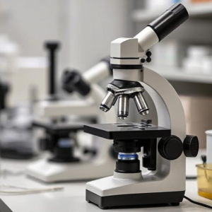
1. Light Microscope (Compound Microscope)
- How it works: A light microscope uses visible light and lenses to magnify small objects. Light passes through the sample, and lenses in the microscope focus the light to create an image you can see.
- Magnification: Usually up to 1000x (this means an object will look 1000 times larger than it is).
- Uses:
- It’s used to view cells, bacteria, and small tissue samples.
- In research, it helps scientists study things like plant cells, bacteria, and even tiny organisms in water.
- Limitations: It can’t magnify very tiny things like viruses or the internal details of cells too clearly, because light can’t reveal finer details at higher magnifications.
2. Electron Microscope
- How it works: Instead of light, an electron microscope uses beams of electrons to magnify objects. Electrons have much shorter wavelengths than light, which means they can reveal much smaller details.
- Types:
- Transmission Electron Microscope (TEM): Sends electrons through a thin sample to create very detailed images of the inside of cells or viruses.
- Scanning Electron Microscope (SEM): Bounces electrons off the surface of the sample to create a 3D image of the outer surface.
- Magnification: Can magnify up to 1,000,000x or more.
- Uses:
- In research, it’s used to look at very tiny things like viruses, bacteria, and the structures inside cells.
- It’s also used to study the surfaces of materials, such as metals or even tiny chips in electronics.
- Limitations: Electron microscopes are very expensive, complex to use, and samples often need to be prepared in special ways, such as being coated with metal or frozen.
3. Fluorescence Microscope
- How it works: A fluorescence microscope uses special lights to excite certain molecules in the sample, making them glow in different colors. This helps researchers see specific structures or molecules inside cells.
- Magnification: Similar to light microscopes, usually up to 1000x, but it can also provide a clearer, more detailed image due to the glowing markers.
- Uses:
- It is widely used in biological and medical research to study specific proteins, cells, and molecules that are tagged with fluorescent markers.
- For example, scientists use it to study the movement of molecules in live cells or to look at different parts of a cell (like the nucleus or mitochondria) that have been specially dyed to glow.
- Limitations: The sample needs to be prepared with fluorescent markers, and the images can sometimes be hard to interpret if the fluorescence is too bright or overlaps.
4. Confocal Microscope
- How it works: A confocal microscope uses lasers and special optics to create very sharp, clear images of thin slices of a sample. It scans the sample point by point and uses a computer to put all the pieces together into one image.
- Magnification: Similar to light and fluorescence microscopes, but it produces sharper, more detailed images, especially for 3D reconstructions.
- Uses:
- It’s commonly used to create detailed images of cells and tissues in 3D, which helps researchers study things like the structure of organs or how cells interact with each other.
- It can also be used to study live cells over time in research.
- Limitations: Like fluorescence microscopes, samples need to be prepared with special fluorescent dyes, and it’s more expensive and complex than a regular light microscope.
5. Scanning Probe Microscope (SPM)
- How it works: SPM uses a tiny probe that scans the surface of a sample to measure its properties, like topography (the surface shape) or electrical activity. It doesn’t use light or electrons like other microscopes but instead uses physical interactions.
- Types:
- Atomic Force Microscope (AFM): Uses a sharp tip to touch the surface of the sample and measures the interaction to create very detailed surface images.
- Scanning Tunneling Microscope (STM): Measures the flow of electrons to map the surface of a material at the atomic level.
- Magnification: It can provide images at the atomic level, revealing incredibly small details.
- Uses:
- Used to study the surface structures of materials at a very fine level. For example, researchers use it to look at atoms or molecules on surfaces or to measure the properties of materials at the nanoscale.
- Limitations: These microscopes are highly specialized and typically used in materials science or nanotechnology research, not for biological samples.
6. Phase Contrast Microscope
- How it works: The phase contrast microscope enhances the contrast of transparent and colorless samples without the need for staining. It uses special optics to amplify differences in light phase as it passes through different parts of the sample.
- Magnification: Similar to light microscopes, up to 1000x or so, but it provides better contrast for certain samples.
- Uses:
- Commonly used to study live cells, especially when researchers don’t want to use stains or dyes that could affect the sample.
- It’s particularly useful for looking at small living organisms, like bacteria or protozoa, in their natural state.
- Limitations: While it increases contrast, it doesn’t provide the high level of detail or color that other types of microscopes can.
Summary of Key Microscopes and Their Uses:
| Microscope Type | How it Works | Best For | Magnification |
|---|---|---|---|
| Light Microscope | Uses visible light and lenses | Viewing cells, bacteria, tissue samples | Up to 1000x |
| Electron Microscope | Uses electron beams to magnify samples | Viewing viruses, cells, small structures | Up to 1,000,000x |
| Fluorescence Microscope | Uses fluorescent dyes to highlight features | Studying specific molecules, proteins | Up to 1000x |
| Confocal Microscope | Uses lasers to create 3D images of thin slices | Detailed imaging of cells and tissues in 3D | Similar to light/fluorescence, but sharper |
| Scanning Probe Microscope | Uses a tiny probe to scan surfaces | Studying atomic-level details of materials | Atomic-level detail |
| Phase Contrast Microscope | Increases contrast in transparent samples | Studying live, unstained cells | Similar to light, with better contrast |
Conclusion:
Microscopes are essential tools in research across many fields like biology, materials science, medicine, and physics. Each type of microscope has a specific job, depending on the size and detail of what you need to see. Whether you’re looking at the structure of cells, the surface of materials, or even atoms, there’s a microscope designed to help you get the best view.


